Liver Disease in Poultry: Causes, Signs, and Prevention
Liver disease in poultry can result from infectious agents, mycotoxins, nutritional imbalance, metabolic disorders, or other causes. Learn how to interpret liver lesions, recognise key signs, use post-mortem findings and laboratory testing, and reduce flock-level risk.
“Liver disease” in poultry is not a single disease.
The liver can be affected by many different groups of causes, including:
- Viruses.
- Bacteria.
- Parasites.
- Mycotoxins.
- Chemicals or toxic substances.
- Nutritional imbalances.
- Metabolic disorders.
- Neoplastic diseases.
The difficulty is that external signs are often non-specific. Chickens may show only:
- Reduced feed intake.
- Poor growth.
- Poor feed conversion ratio (FCR).
- Reduced egg production.
- Depression.
- Increased mortality.
At the same time, findings such as a yellow liver, enlarged liver, distended gallbladder, or green droppings are not sufficient to determine the cause.
A more appropriate approach is therefore to combine:
flock age → mortality pattern → feed quality → post-mortem lesions → lesions in other organs → appropriate laboratory testing.
Abnormal liver findings at post-mortem: 5 directions to investigate
| Observed lesion | What should be investigated further |
| Enlarged, pale, or yellow liver | Fat accumulation, FLHS, aflatoxin exposure, or certain infectious diseases |
| Friable liver, haemorrhage, or blood in the abdominal cavity | FLHS or other causes of liver damage and haemorrhage |
| Multiple white, yellow, or grey necrotic foci | Infectious disease, histomoniasis, or systemic bacterial infection depending on the overall disease pattern |
| Multiple nodules or tumours | Marek’s disease, avian leukosis, or another neoplastic disease |
| Abnormal liver with rapidly increasing mortality | Conduct a complete post-mortem examination, review flock age, feed and vaccination history, and collect samples early where appropriate |
The cause should not be diagnosed from liver colour alone. Flock age, mortality pattern, lesions in other organs, feed history, and appropriate testing are needed to narrow the differential diagnosis.
1. An abnormal liver does not automatically mean a primary “liver disease”
The liver performs many important functions, including:
- Carbohydrate metabolism.
- Lipid metabolism.
- Protein synthesis.
- Bile production.
- Storage of selected nutrients.
- Metabolism and elimination of many substances.
- Regulation of processes involved in growth and reproduction.
When the liver is affected, flock performance may include:
- Reduced weight gain.
- Poor FCR.
- Reduced egg production.
- Reduced egg quality.
- Poorer overall health.
- Increased mortality.
However, liver lesions should be considered within several different categories.
Hepatitis
Hepatitis is inflammation of liver tissue and may be associated with:
- Viruses.
- Bacteria.
- Parasites.
- Certain toxic substances.
Fatty liver
Fatty liver refers to abnormal lipid accumulation within hepatocytes.
It may be associated with:
- Prolonged positive energy balance.
- Metabolic disorders.
- High egg-production periods.
- Certain toxins.
Toxic liver injury
Certain mycotoxins or chemicals may cause:
- Hepatocellular necrosis.
- Lipid accumulation.
- Haemorrhage.
- Reduced liver function.
Hepatic tumours
Marek’s disease and avian leukosis may produce tumours or abnormal cellular infiltration in the liver.
These cases should not simply be grouped under the term “hepatitis.”
2. Using liver lesions to guide the differential diagnosis
Post-mortem examination is valuable because it helps move from non-specific clinical signs to more specific lesion patterns.
However, each lesion is only suggestive, not a final diagnosis.
Enlarged liver
Hepatomegaly may occur with:
- Inflammation.
- Septicaemia.
- Fatty liver.
- Neoplastic disease.
- Certain viral diseases.
If only the liver is examined, identifying the cause can be difficult.
Other organs should also be assessed, including:
- Spleen.
- Kidneys.
- Bursa of Fabricius.
- Heart.
- Lungs.
- Air sacs.
- Intestines.
- Caeca.
Yellow or pale liver
This may be associated with:
- Lipid accumulation.
- Aflatoxicosis.
- FLHS.
- Certain infectious diseases.
However, liver colour may also be influenced by:
- Age.
- Diet.
- Pigments.
For example, chickens receiving diets rich in xanthophyll may have a more yellow liver without necessarily having FLHS.
Haemorrhagic liver
Possible findings include:
- Petechial haemorrhages.
- Larger areas of haemorrhage.
- Liver rupture.
- Blood clots in the abdominal cavity.
Possible causes vary widely and may include:
- FLHS.
- Infectious disease.
- Toxicosis.
- Other forms of injury.
Body condition, flock age, and mortality pattern should therefore also be evaluated.
Necrotic foci
White, yellow, or grey foci should be assessed according to:
- Number.
- Size.
- Shape.
- Distribution.
- Whether similar lesions occur in other organs.
For example, in histomoniasis in turkeys, liver lesions should be assessed together with the caeca.
Tumours
If the liver is:
- Affected by multiple nodules.
- Markedly enlarged.
- Affected by localised or diffuse masses.
and lesions are also found in:
- Spleen.
- Kidneys.
- Bursa of Fabricius.
- Other organs.
neoplastic diseases such as:
- Marek’s disease.
- Avian leukosis.
should be included in the differential diagnosis.
Distended gallbladder
A distended gallbladder may occur simply when poultry:
- Reduce feed intake.
- Stop eating for a period.
Therefore:
Gallbladder distension alone is not evidence of liver disease.
3. When mortality rises rapidly in a young flock: which infectious diseases should be prioritised?
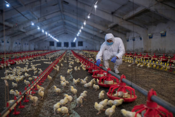
Flock age is particularly important when interpreting liver lesions.
Some infectious diseases are most relevant in specific age groups or poultry species.
Fowl adenovirus and inclusion body hepatitis
Inclusion Body Hepatitis (IBH) is associated with certain fowl adenoviruses (FAdV) belonging to the genus Aviadenovirus.
It is particularly important in young broiler chickens.
Possible findings include:
- Sudden increases in mortality.
- Depression.
- Ruffled feathers.
- Huddling.
- Yellow-green droppings.
- Enlarged liver.
- Pale or yellow liver.
- Petechial haemorrhages.
- Areas of necrosis.
IBH and FAdV-associated hepatitis–hydropericardium syndrome are often particularly relevant in chickens under approximately six weeks of age, although age distribution can vary according to:
- Viral strain.
- Flock status.
- Epidemiological factors.
Diagnosis should integrate:
- Flock age.
- Mortality pattern.
- Lesions.
- Histopathology.
- PCR where appropriate.
FAdV detection alone should not automatically be interpreted as confirmation of IBH because adenoviruses may also be detected in some birds without compatible clinical disease.
Duck viral hepatitis
Duck viral hepatitis is an acute disease of particular importance in ducklings, especially during the first few weeks of life.
It may cause:
- Rapid death.
- High mortality.
- Liver lesions.
- Certain neurological signs before death.
The term “duck hepatitis” should not be used as a general diagnosis for liver disease in all poultry species.
Septicaemia
Several bacterial pathogens may cause liver lesions as part of systemic infection, including:
- Salmonella spp.
- Escherichia coli.
- Pasteurella multocida.
In these situations, the liver is only one part of the disease process.
Other organs should also be examined, including:
- Spleen.
- Heart.
- Lungs.
- Air sacs.
- Intestines.
- Other organs.
An abnormal liver alone should not be used to conclude that a pathogen is specifically causing “liver disease.”
Histomoniasis
Histomonas meleagridis causes histomoniasis, commonly known as blackhead disease.
The disease is particularly severe in turkeys.
Findings may include:
- Severe caecal lesions.
- Characteristic necrotic lesions in the liver.
- High mortality.
Chickens can carry the organism and may have milder disease, although clinical disease can also occur.
When liver lesions raise suspicion of histomoniasis, the caeca should also be examined rather than focusing only on the liver.
Marek’s disease and Gumboro disease
Marek’s disease may cause:
- Enlarged liver.
- Tumours.
- Neoplastic cellular infiltration.
However, it is a viral neoplastic disease rather than primary hepatitis.
Gumboro disease, or infectious bursal disease, primarily affects:
- The bursa of Fabricius.
- The immune system.
It can increase susceptibility to secondary infection but should not be considered a typical primary cause of hepatitis.
4. When feed is suspected: how can mycotoxin exposure be differentiated from infectious disease?
Mycotoxins are toxic compounds produced by certain moulds in feed ingredients or finished feed.
Risk may increase when:
- Ingredients have high moisture content.
- Storage conditions are hot and humid.
- The feed store leaks.
- Feed is stored for prolonged periods.
- Ingredient management is poor.
However, detecting damp or mouldy feed does not automatically prove that mycotoxins are causing the disease problem.
Aflatoxin
Aflatoxin is one of the most important hepatotoxic mycotoxins in poultry.
Possible effects include:
- Hepatocellular damage.
- Necrosis.
- Lipid accumulation.
- Haemorrhage.
- Reduced protein synthesis.
- Immunosuppression.
- Reduced weight gain.
- Poor FCR.
- Reduced egg production.
Clinical expression may be acute or chronic depending on:
- Exposure dose.
- Duration of exposure.
- Species.
- Age.
- Flock health status.
Ochratoxin and other mycotoxins
Not all mycotoxins have the same primary target organ.
Ochratoxin is particularly associated with:
- Nephrotoxicity.
- Immunosuppression.
although it may also affect the liver and other organs.
Other groups, including:
- Fumonisins.
- Trichothecenes.
have different toxicity profiles.
It is therefore not possible to identify the exact mycotoxin simply from liver lesions.
What should be done first when feed is suspected?
A practical sequence is:
- Identify the feed batch currently being used.
- Isolate or discontinue the suspect batch where the level of concern justifies doing so.
- Inspect feed-storage conditions.
- Inspect ingredients.
- Collect representative samples.
- Record supplier and delivery information.
- Submit samples for quantitative testing where required.
Feed that does not visibly appear mouldy may still contain mycotoxins.
Conversely, visible mould does not reveal the exact toxin or its concentration.
Toxin binders are not a solution for every situation
The effectiveness of a toxin binder depends on:
- Mycotoxin type.
- Product composition.
- Inclusion rate.
- Feed composition.
- Level of exposure.
Some products may bind aflatoxin more effectively, while performance against other mycotoxins may be lower.
Therefore:
A toxin binder should not be used as justification for continuing to feed a batch that is suspected of posing a mycotoxin risk.
5. In laying hens: when should FLHS be considered?
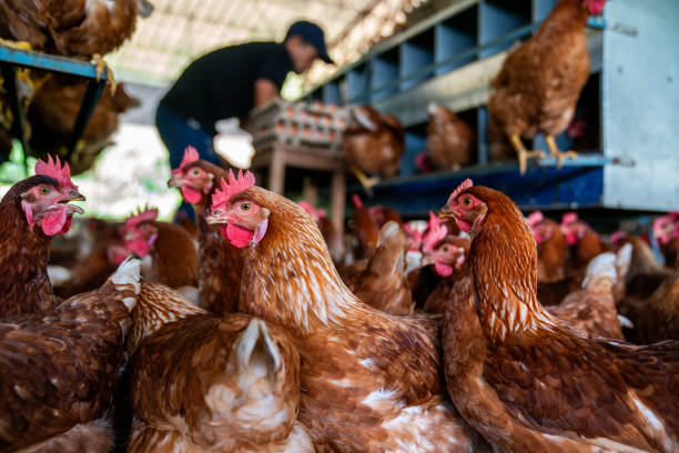
Fatty Liver Haemorrhagic Syndrome (FLHS) is an important metabolic disorder in laying hens.
Risk is often more relevant in:
- High-producing hens.
- Over-conditioned birds.
- Flocks with prolonged positive energy balance.
- Hot-weather periods.
Some affected hens may die suddenly without obvious previous clinical signs.
Lesions that may raise suspicion of FLHS
A combination may include:
- Enlarged liver.
- Pale liver.
- Very friable liver.
- Petechial or larger haemorrhages.
- Blood clots in the abdominal cavity.
- Excess abdominal fat.
In particular, a friable haemorrhagic liver with blood in the abdominal cavity in laying hens is more suggestive of FLHS than liver colour alone.
A yellow liver does not automatically mean FLHS
A yellow liver may also be related to:
- Diet.
- Pigmentation.
- Aflatoxin exposure.
- Other forms of lipid accumulation.
Assessment should therefore also consider:
- Flock age.
- Stage of lay.
- Body condition.
- Abdominal fat.
- Haemorrhagic lesions.
- Diet.
Role of nutrition
Fatty liver can be associated with an imbalance between:
- Lipid synthesis.
- Lipid transport.
- Lipid utilisation.
Risk may increase where:
- Energy intake exceeds requirements.
- Physical activity is low.
- Metabolic demand is high during lay.
- The ration is imbalanced.
- Metabolic stress is present.
Energy intake should not be increased excessively simply to stimulate egg production.
Methionine, choline, betaine, vitamin E, and selenium
These nutrients all participate in important metabolic processes.
However:
- Methionine.
- Choline.
- Betaine.
- Vitamin E.
- Selenium.
should not be treated as general “liver medicines” when the basal diet already meets nutritional requirements.
Supplementation should be based on:
- Feed formulation.
- Ingredients.
- Nutrient requirements.
- Production stage.
- Professional nutritional assessment.
6. External signs that can easily be misinterpreted
One of the difficulties with liver disorders is that clinical signs are often highly non-specific.
Green or yellow droppings
Green droppings may occur when chickens:
- Eat less.
- Stop eating.
- Have systemic disease.
Yellow or loose droppings may also be associated with:
- Intestinal disease.
- Infection.
- Feed changes.
Therefore:
Dropping colour alone is not sufficient to diagnose liver disease.
Pale or cyanotic comb
This may be associated with:
- Anaemia.
- Blood loss.
- Cardiopulmonary disease.
- Systemic illness.
- Severe haemorrhage associated with FLHS.
It is not a liver-specific sign.
Reduced egg production
Liver dysfunction may affect production, but lower egg output may also result from:
- Stress.
- Infectious disease.
- Nutrition.
- Lighting.
- Flock age.
Poor weight gain and poor FCR
These may result from many problems, including:
- Nutritional imbalance.
- Intestinal disease.
- Respiratory disease.
- Infection.
- Mycotoxin exposure.
- Metabolic disorders.
Production indicators are therefore most useful when interpreted as trends over time, rather than as standalone evidence of liver disease.
7. When should post-mortem examination and laboratory testing be performed?
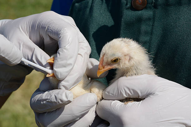
A fixed rule such as “wait 3–5 days if there is no improvement” should not be followed when mortality is already increasing or unusual lesions are present.
Post-mortem examination and early sampling should be considered when:
- Mortality exceeds expected levels.
- Mortality increases rapidly.
- Multiple birds die with similar lesions.
- Marked liver haemorrhage is present.
- Multiple necrotic liver lesions are found.
- IBH is suspected.
- Mycotoxin exposure is suspected.
- Septicaemia is suspected.
- Multiple internal tumours are present.
- The initial diagnosis does not match the lesions.
- Current treatment or management lacks a clear diagnostic basis.
- Antimicrobial selection needs laboratory support.
Which birds should be selected for post-mortem examination?
Priority should generally be given to:
- Birds showing typical clinical signs.
- Recently dead birds.
Carcasses that have been dead for too long may develop:
- Post-mortem changes.
- Secondary contamination.
which can reduce diagnostic value.
Do not examine only the liver
A suspected liver case should still include examination of:
- Spleen.
- Kidneys.
- Bursa of Fabricius.
- Intestines.
- Caeca.
- Heart.
- Air sacs.
- Other organs.
This is particularly important for differentiating:
- Systemic infectious diseases.
- Neoplastic disease.
- Histomoniasis.
- Secondary causes of liver lesions.
8. Collect samples according to the diagnostic question — not for a generic “liver test”
There is no single sample or storage method suitable for every diagnostic test.
The first question should be:
What cause is suspected, and what diagnostic question should the laboratory answer?
Histopathology
Histopathology may help evaluate:
- Necrosis.
- Inflammation.
- Inclusion bodies.
- Neoplasia.
- Other cellular changes.
Samples should include:
- Lesion tissue.
- A portion of relatively normal adjacent tissue where appropriate.
Samples should be fixed according to the diagnostic laboratory’s instructions.
PCR or virology
Samples should be selected according to the suspected pathogen.
Depending on the disease, samples may include:
- Liver.
- Spleen.
- Other relevant tissues.
A generic “liver PCR panel” should not be requested without a defined diagnostic suspicion.
Bacteriology
Where bacterial infection is suspected:
- Collect samples aseptically.
- Prioritise clinically affected or recently dead birds.
- Where practical, collect samples before antimicrobial treatment.
Samples may be used for:
- Bacterial isolation.
- Identification.
- Antimicrobial susceptibility testing.
Mycotoxin testing
Where mycotoxin exposure is suspected, important samples generally include:
- Finished feed.
- Suspect feed ingredients.
Samples should be representative of the relevant batch.
Records should include:
- Batch number.
- Delivery date.
- Supplier.
- Date feeding began.
Taking a single handful of feed from one location in the store may not represent the entire batch.
9. Preventing liver problems at flock level requires multiple control points
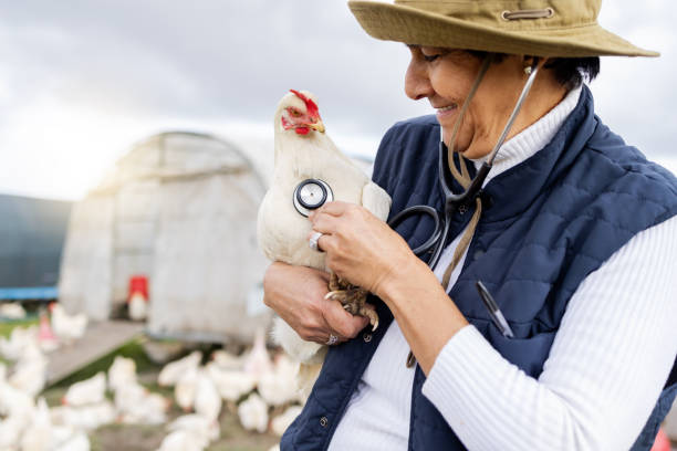
No single product or intervention can prevent every cause of liver disease.
Prevention requires several complementary measures.
Breeding-source control
Priority should be given to sources that:
- Are traceable.
- Have clear flock-health programmes.
- Are suitable for the production system.
Disease-specific vaccination
There is no general “liver disease vaccine.”
For example:
- Marek’s disease has specific vaccines.
- Gumboro disease has specific vaccines.
- FAdV vaccines are used in some countries and production programmes to control IBH/HHS.
Actual use depends on:
- Vaccine registration status.
- Epidemiological risk.
- Relevant serotype.
- Breeder-flock programme.
- Veterinary advice.
Feed management
Feed should not simply continue to be used without further assessment when it:
- Has an abnormal odour.
- Is damp.
- Is caked.
- Is clearly spoiled.
- Shows substantial mould growth.
Feed storage should be:
- Dry.
- Well ventilated.
- Protected from leaks.
Feed should be stored appropriately off the floor, with effective insect and rodent control.
Retaining feed samples
Keeping representative samples from each feed batch can be very useful.
This supports:
- Traceability.
- Comparison across production periods.
- Laboratory testing if a problem develops.
Water management
Farms should monitor:
- Water source.
- Storage tanks.
- Biofilm in pipelines.
- Drinkers or nipple systems.
Contaminated water can contribute to multiple health problems even when it is not a specific cause of liver disease.
Nutrition
Diets should be properly balanced from the outset rather than relying on “liver supplements” after problems appear.
Monitoring should include:
- Energy.
- Protein.
- Amino acids.
- Vitamins.
- Minerals.
- Flock body weight.
Heat stress and environment
Heat stress is not a common direct cause of hepatitis, but it can influence:
- Feed intake.
- Energy metabolism.
- Oxidative stress.
- Gut health.
- Immunity.
In laying hens, hot weather may also contribute to FLHS risk where metabolic predisposing factors are already present.
Biosecurity
Basic principles include:
- Control of people.
- Control of vehicles.
- Farm zoning.
- Dedicated equipment where appropriate.
- Cleaning before disinfection.
- Wild-bird control.
- Rodent control.
- Appropriate carcass disposal.
10. Practical scenario: 4–5-week-old broilers with enlarged, pale, haemorrhagic livers
Consider a broiler flock approximately 4–5 weeks of age showing:
- Reduced feed intake.
- Depression.
- Yellow-green droppings.
- Increasing mortality.
Post-mortem examination identifies:
- Enlarged liver.
- Pale liver.
- Petechial haemorrhages.
At the same time, the farm identifies a batch of feed ingredients that appears:
- Damp.
- Poorly stored.
Can mycotoxicosis be diagnosed immediately?
No.
The feed history increases suspicion but does not establish the diagnosis.
Similar lesions may occur in:
- IBH.
- Septicaemia.
- Certain toxicoses.
- Other systemic diseases.
What sequence should be considered?
A reasonable approach may include:
- Identify and reassess the relevant feed batch.
- Isolate the suspect source where necessary.
- Conduct post-mortem examinations on additional representative birds.
- Examine all relevant organs.
- Consider feed testing.
- Collect tissues for pathogen testing where the disease pattern suggests it.
- Monitor flock response after changing the feed source.
If the flock improves after the feed is changed, this provides supporting evidence for the hypothesis.
However, improvement alone does not prove mycotoxicosis in the absence of:
- Laboratory testing.
- Compatible lesions.
- Sufficient epidemiological evidence.
Frequently asked questions about liver disease in poultry
Is liver disease contagious between birds?
It depends on the cause.
Where the cause is:
- Viral.
- Bacterial.
- Another infectious agent.
the pathogen may spread through the flock.
Where liver injury is associated with:
- Mycotoxins.
- Diet.
- Chemicals.
- Metabolic disorders.
the condition itself is not contagious.
However, many birds may still be affected simultaneously because they share the same feed source or environmental exposure.
Does a yellow liver always mean fatty liver?
No.
A yellow liver may be associated with lipid accumulation, but it can also be influenced by:
- Diet.
- Pigments.
- Age.
- Certain diseases.
Additional findings should be assessed, including:
- Liver friability.
- Haemorrhage.
- Abdominal fat.
- Body condition.
- Other lesions.
Are green droppings a sign of liver disease?
Not necessarily.
Green droppings commonly occur when chickens:
- Reduce feed intake.
- Stop eating.
- Develop systemic illness.
Dropping colour is meaningful only when interpreted alongside:
- Flock age.
- Lesions.
- Feed intake.
- Mortality.
- Other clinical signs.
Is FLHS an infectious disease?
No.
FLHS is a metabolic disorder, primarily affecting laying hens, and may be influenced by:
- Energy balance.
- Sex.
- Hormonal factors.
- Activity level.
- Environmental temperature.
Should “liver supplements” be used routinely?
Liver-support products should not be regarded as substitutes for:
- A balanced diet.
- Mycotoxin control.
- Appropriate treatment of infectious diseases.
- Removal of toxin sources.
- Environmental management.
Supplement use should be based on a defined nutritional or management objective.
Can toxin binders prevent all mycotoxins?
No.
Effectiveness depends heavily on:
- Toxin type.
- Binder composition.
- Inclusion rate.
- Diet composition.
Binders do not replace:
- Ingredient-quality control.
- Appropriate storage.
- Removal of high-risk feed batches.
A Multi-Cause Approach to Poultry Liver Health at VIETSTOCK 2026
Liver problems in poultry may result from infectious disease, mycotoxins, nutritional imbalance, or metabolic disorders. For this reason, liver colour or a single external sign is rarely sufficient to determine the cause. Combining flock data, feed quality, post-mortem findings, and appropriate laboratory testing helps farms make more accurate decisions and avoid continuing interventions that do not address the underlying problem.
At VIETSTOCK 2026 – Vietnam’s Premier International Feed, Livestock & Meat Industry Show, poultry-health challenges are addressed alongside other production factors such as nutrition, feed, veterinary medicine, and housing conditions. The event is expected to bring together 300 brands, over 10,000 sqm of exhibition area, and 13,000 trade visitors from 40 countries.
For farms seeking to identify causes and reduce liver-health risks at flock level, participants can explore solutions in areas such as:
- Feed, ingredients, and additives: solutions that support ingredient-quality control, nutrition, and factors affecting liver health.
- Mycotoxin control: approaches that support assessment and reduction of mycotoxin risk in animal feed.
- Veterinary diagnostics: technologies that support pathogen identification, lesion assessment, and selection of appropriate diagnostic tests.
- Poultry health and disease prevention: developments in vaccination, biosecurity, and infectious-disease control.
- Production-monitoring technology: tools that support monitoring of feed intake, productivity, mortality, and abnormal flock-level trends.
The One Health Forum on 22 October 2026 also includes the session “Mycotoxin management and its One Health implications.” This topic is directly relevant to one of the important causes of liver injury in poultry and helps participants view mycotoxin risk not only in terms of flock performance, but also in relation to feed quality, animal health, and production-system sustainability.
Date: 21–23 October 2026
Venue: Saigon Exhibition and Convention Center (SECC), 799 Nguyen Van Linh Street, Tan My Ward, Ho Chi Minh City, Vietnam
Event website: https://www.vietstock.org/en/
👉 VIETSTOCK 2026 Visitor Registration Information
👉 Learn more about the One Health Forum
Contact information:
Exhibiting: Ms. Sophie Nguyen – [email protected]
Visitor Support: Ms. Phuong – [email protected]
Marcom Support: Ms. Anita Pham – [email protected]