Avian Leukosis in Poultry: Signs, Transmission, and Prevention
Avian leukosis is a group of neoplastic diseases caused by Avian Leukosis Virus (ALV), primarily affecting chickens. Learn about its clinical signs, vertical and horizontal transmission, tumour lesions, differential diagnosis with Marek’s disease, and prevention strategies focused on breeder-flock control.
Avian leukosis is a group of neoplastic diseases caused by Avian Leukosis Virus (ALV). Chickens are the natural host and the species of greatest importance in commercial poultry production.
Unlike some acute infectious diseases, avian leukosis does not usually cause sudden mass mortality over a short period. Its impact often develops more gradually through cumulative mortality, reduced weight gain, lower egg production, and increased culling rates.
One of the most important epidemiological characteristics of ALV is its ability to spread vertically from infected hens to their offspring through the egg. Disease control must therefore focus primarily on breeder-flock and chick-source health status rather than only removing birds that have already developed tumours.
There is currently no specific treatment or widely used effective vaccine for controlling avian leukosis.
4 key points to remember about avian leukosis
- The disease often develops gradually: Avian leukosis does not usually cause sudden mass mortality. Instead, mortality may rise progressively, while birds become weak, show poorer weight gain, reduced egg production, and higher culling rates. Lymphoid leukosis is generally more apparent in chickens from approximately 14–16 weeks of age onward.
- Breeding stock is a critical control point: ALV can be transmitted from infected hens to offspring through the egg. Chickens infected during embryonic development may carry and shed the virus for prolonged periods without obvious clinical signs.
- The presence of tumours alone does not confirm avian leukosis: Differential diagnosis should include Marek’s disease, reticuloendotheliosis, and other neoplastic diseases. A positive PCR result alone is also not sufficient to establish ALV as the cause of a tumour.
- Control should begin with breeder flocks: There is currently no specific treatment or widely used effective vaccine for ALV. Prevention relies mainly on controlled breeding sources, reduction of vertical transmission, biosecurity, and monitoring of flock-performance data.
1. What is avian leukosis?
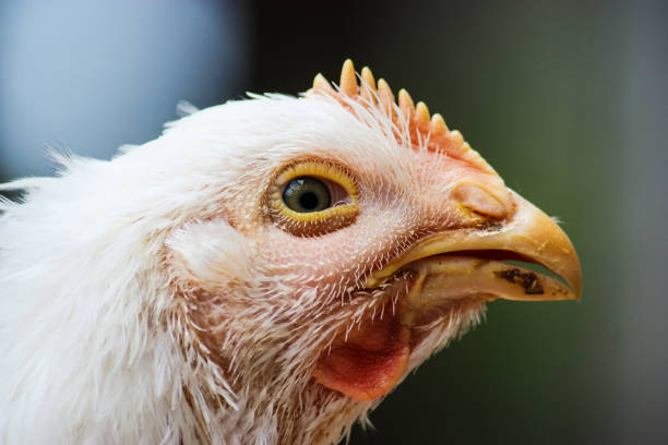
Avian leukosis is a group of neoplastic diseases caused by viruses belonging to the avian leukosis/sarcoma virus group.
ALV belongs to:
- The family Retroviridae.
- The genus Alpharetrovirus.
The virus can integrate its genetic material into host cells. Under certain conditions, this process may promote abnormal cellular transformation and proliferation, eventually resulting in tumour formation.
Disease expression depends on multiple factors, including:
- Viral subgroup.
- Chicken line.
- Age at infection.
- Genetic susceptibility.
- Immune status.
- Route of infection.
Chickens are the most extensively documented and studied host.
Other bird species may carry related viruses, but in commercial poultry production, the economic importance of avian leukosis is primarily associated with chickens.
2. Common forms of avian leukosis
2.1. Lymphoid leukosis
This is the classical and most commonly discussed form of avian leukosis.
Tumours originate mainly from B lymphocytes and are commonly associated with the bursa of Fabricius.
Tumours are generally not obvious when birds are very young.
Lymphoid leukosis is more commonly recognised in chickens:
- From approximately 14–16 weeks of age onward.
- Near or after sexual maturity.
Tumours may occur in:
- Bursa of Fabricius.
- Liver.
- Spleen.
- Kidneys.
- Other organs.
2.2. Myeloid leukosis
Myeloid leukosis involves abnormal proliferation of cells of the myeloid lineage and has been particularly associated with certain ALV infections, with ALV subgroup J being of particular importance.
Lesions may occur in:
- Liver.
- Spleen.
- Bone marrow.
- Bone.
- Other tissues or organs.
2.3. Less common forms
Other neoplastic conditions within the leukosis/sarcoma group may include:
- Erythroblastosis.
- Myelocytomatosis.
- Certain forms of sarcoma.
The disease form cannot be classified reliably from external appearance alone.
Where confirmation is required, diagnosis should combine:
- Post-mortem examination.
- Histopathology.
- Appropriate laboratory testing.
3. Common clinical signs and lesions
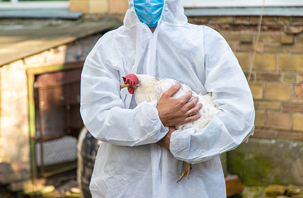
3.1. External signs
Affected chickens may show:
- Progressive weight loss.
- Reduced feed intake.
- Wasting.
- Pale comb and wattles.
- Abdominal enlargement.
- Poor feather condition.
- Reduced activity.
- Poor weight gain.
- Reduced or absent egg production.
These signs usually develop gradually and are not specific to ALV.
At flock level, farmers may notice:
- Mortality increasing gradually over several weeks.
- Reduced egg production.
- Poorer growth performance.
- Increased culling.
- Flock productivity below expected levels.
3.2. Liver and spleen
The liver may be:
- Enlarged.
- Affected by multiple white or greyish nodules or masses.
- Involved in diffuse or localised neoplastic lesions.
The spleen may be:
- Enlarged.
- Structurally abnormal.
- Affected by infiltrative areas or tumours.
3.3. Kidneys and other organs
The kidneys may:
- Become enlarged.
- Be infiltrated by neoplastic cells.
- Develop abnormal masses.
In rare cases, a large renal tumour may cause compression and result in abnormal movement or locomotor signs.
However, peripheral nerve lesions are not a typical feature of lymphoid leukosis.
3.4. Bursa of Fabricius
In lymphoid leukosis, the bursa of Fabricius is diagnostically important.
Possible findings include:
- Abnormal enlargement.
- Altered follicles.
- Nodular or diffuse tumours.
However, gross examination of the bursa alone is not sufficient to confirm the disease.
3.5. Ovaries
In laying hens, tumours or neoplastic infiltration may involve the ovaries and contribute to reduced reproductive performance.
4. How is avian leukosis transmitted?
4.1. Vertical transmission through the egg
This is one of the most important epidemiological routes of ALV transmission.
An infected hen may transmit the virus through the egg to the developing embryo.
Chicks infected during embryonic development may:
- Carry the virus from hatch.
- Remain clinically normal for prolonged periods.
- Develop persistent viraemia.
- Shed virus.
- Become a source of infection for other birds.
Congenitally infected chickens may fail to develop an effective neutralising antibody response because of immune tolerance.
This is why ALV control must begin with parent flocks and breeding establishments.
4.2. Horizontal transmission
ALV can also spread between birds after hatching.
Horizontal transmission is particularly relevant when young chicks are exposed early to:
- Infected or virus-shedding birds.
- Virus-containing secretions or excretions.
- Contaminated equipment.
However, ALV is less stable outside the host than several other important poultry viruses and can be relatively readily inactivated through appropriate cleaning and disinfection.
The farm environment therefore contributes to horizontal transmission, but it should not be regarded as a more important risk source than infected breeding stock.
4.3. Equipment and husbandry procedures
Shared equipment may contribute to pathogen transmission if it is not properly cleaned.
Particular attention should be paid to:
- Husbandry equipment.
- Feeders and drinkers.
- Veterinary equipment.
- Needles.
Avoiding reuse of needles between multiple birds remains an appropriate biosecurity principle, even though this is not considered the primary transmission route for ALV.
5. Risk factors and the importance of chick source
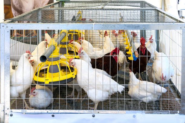
5.1. Chick sources without an ALV control programme
This should be one of the first risk factors assessed.
Farms should avoid or carefully evaluate chicks from:
- Untraceable sources.
- Breeding establishments without a clear ALV-control programme.
- Parent flocks with a history of tumours or abnormal productivity that has not been investigated.
With ALV, a chicken that appears healthy is not necessarily free of infection.
5.2. Congenitally infected chicks
Chicks infected through the egg may become long-term virus shedders.
Removing only birds that develop tumours later in life is therefore insufficient to eliminate ALV from a flock when the original source of infection is the breeding population.
5.3. Horizontal spread within the flock
Conditions that increase contact between birds may facilitate horizontal transmission, including:
- Mixing birds from multiple sources.
- Sharing equipment.
- Inadequate cleaning between areas.
- Uncontrolled mixing of flocks.
Stocking density, ventilation, and stress may affect general flock health and management, but they should not be described as specific causes of avian leukosis.
5.4. Failure to monitor flock data
ALV-associated losses may develop gradually.
Farms should monitor:
- Weekly mortality.
- Egg production.
- Weight gain.
- Culling rate.
- Post-mortem findings.
Long-term trends over several weeks are more informative than observations from only a few affected birds.
6. Prevention and control of ALV
6.1. Control at breeder-flock level
The most important strategy is reducing the introduction of virus through breeding stock.
Priority should be given to chicks from systems with:
- Traceable parent flocks.
- ALV surveillance programmes.
- Virus-eradication or control programmes.
- Appropriate breeder-flock health records.
In more specialised breeding programmes, testing methods may include:
- Virus detection.
- Antigen testing.
- Detection of p27 antigen in egg albumen.
- Other virological or molecular techniques.
The objective is to identify hens that are shedding virus and reduce transmission to offspring.
6.2. Management of newly introduced flocks
Separation and monitoring of newly introduced birds remains useful within an overall biosecurity programme, but it does not replace breeder-source control.
During the observation period:
- Keep new birds separated.
- Use dedicated equipment.
- Monitor mortality.
- Record abnormal signs.
- Review source documentation.
6.3. Cleaning and disinfection
Between production cycles:
- Remove manure and litter.
- Dispose of poultry carcasses appropriately.
- Clean feeders and drinkers.
- Clean housing surfaces.
- Apply an appropriate disinfectant.
- Clean equipment.
- Control movement of people and equipment between areas.
ALV can be relatively readily inactivated outside the host when cleaning and disinfection are performed correctly.
6.4. Record management
Farms should retain records of:
- Chick source.
- Incoming batch.
- Date of placement.
- Mortality.
- Production.
- Post-mortem findings.
- Laboratory results where available.
If multiple batches from the same source develop similar problems, these records can be important for tracing the risk.
7. What should be done when avian leukosis is suspected?
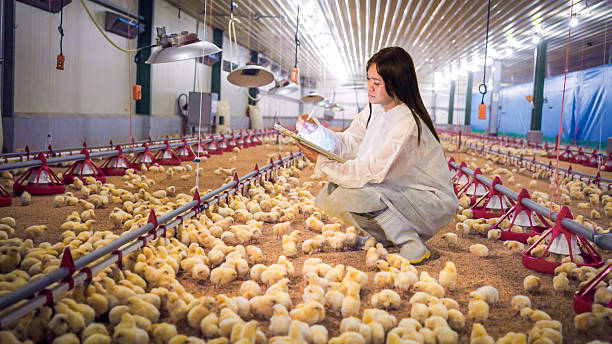
Attention should not focus only on a few visibly affected birds.
ALV may be present in birds without obvious clinical disease.
When chickens show:
- Progressive wasting.
- Abdominal enlargement.
- Pale combs.
- Reduced egg production.
- Internal tumours at post-mortem examination.
the following steps should be considered.
Step 1: Record and assess the whole flock
Review:
- Flock age.
- Chick source.
- Mortality rate.
- Mortality trends over time.
- Production performance.
- Weight gain.
- Number of birds with neoplastic lesions.
Step 2: Manage affected birds
Weak birds or birds selected for sampling may be separated to facilitate monitoring and examination.
However, isolating a few affected birds does not mean ALV has been controlled within the flock.
Step 3: Conduct post-mortem examinations on appropriate birds
Record lesions involving:
- Liver.
- Spleen.
- Kidneys.
- Bursa of Fabricius.
- Reproductive organs.
- Peripheral nerves.
Step 4: Contact a veterinarian or diagnostic laboratory
Professional investigation is particularly appropriate when:
- Mortality continues to rise.
- Multiple visceral tumours are found.
- ALV cannot be distinguished from Marek’s disease.
- Similar problems occur in consecutive flocks.
- Abnormalities are suspected in breeding stock.
Step 5: Review the breeding source
If ALV is confirmed or strongly suspected, review:
- Chick source.
- Parent flock.
- Other batches from the same source.
- History of ALV control.
8. Diagnosis and sample collection

8.1. Do not diagnose avian leukosis by PCR alone
This is particularly important.
ALV may be detected in chickens that do not have tumours caused by avian leukosis.
Therefore:
A positive PCR result or detection of ALV alone is not sufficient to confirm that a tumour is lymphoid leukosis.
Diagnosis should integrate:
- Flock age.
- Clinical signs.
- Gross lesions.
- Histopathology.
- Virological or molecular testing where appropriate.
8.2. Role of histopathology
Histopathology is highly valuable for:
- Confirming the nature of the tumour.
- Differentiating types of lymphoma.
- Supporting differentiation between avian leukosis, Marek’s disease, and other neoplastic conditions.
In lymphoid leukosis, neoplastic cells are generally relatively uniform and predominantly of B-cell origin.
8.3. Common samples
Depending on the diagnostic objective, samples may include:
- Tumour tissue.
- Liver.
- Spleen.
- Bursa of Fabricius.
- Kidney.
- Other abnormal tissues.
The diagnostic laboratory should be contacted before sample collection because:
- Histopathology samples usually require fixation.
- Samples for virus detection or PCR may require different storage conditions.
All samples should not automatically be placed into the same preservative before the intended test has been determined.
9. Differentiating avian leukosis from Marek’s disease and other neoplastic diseases
Marek’s disease is one of the most important differential diagnoses because both diseases can cause visceral tumours.
| Feature | Avian leukosis – lymphoid leukosis | Marek’s disease |
| Typical age | Usually over 14–16 weeks | May occur from approximately 3 weeks of age |
| Neoplastic cell origin | Predominantly B lymphocytes | Predominantly T lymphocytes |
| Bursa of Fabricius | Often enlarged and may contain tumours | Often atrophied; bursal tumours may still occur |
| Peripheral nerves | Usually not enlarged | Nerve enlargement is an important finding |
| Leg/wing paralysis | Not typical | More common |
| Skin/muscle tumours | Less typical | May occur |
| Vaccine | No widely used effective vaccine | Effective protective vaccines are available |
This table is intended only to support differential diagnosis and does not replace professional diagnostic investigation.
In particular:
- Tumours in the bursa of Fabricius may support lymphoid leukosis but do not completely exclude Marek’s disease.
- Enlargement of peripheral nerves strongly supports Marek’s disease.
- Marek’s disease may occur in substantially younger birds than lymphoid leukosis.
In addition to Marek’s disease, reticuloendotheliosis should also be considered in some cases because it can produce similar lymphoid tumours.
When lesions are atypical, histopathology and specialised laboratory testing are preferable to diagnosis based solely on gross post-mortem findings.
10. Is there treatment, a vaccine, or a risk to humans?
10.1. Is there a treatment for ALV?
There is currently no specific treatment that eliminates ALV from infected chickens.
Antibiotics:
- Do not kill ALV.
- Do not make tumours disappear.
- Should not be used simply because avian leukosis is suspected.
Antibiotics may only be considered by a veterinarian where there is an appropriate secondary bacterial infection.
Disease management instead focuses on:
- Breeding-source control.
- Flock monitoring.
- Diagnostic confirmation.
- Management of affected birds.
- Reduction of horizontal transmission.
- Management of infected breeder flocks.
10.2. Is there a vaccine against avian leukosis?
There is currently no widely used effective vaccine for controlling ALV comparable with vaccination against Marek’s disease.
Control therefore depends mainly on:
- Eliminating or reducing virus in breeder flocks.
- Controlling vertical transmission.
- Selecting monitored breeding sources.
- Biosecurity.
- Epidemiological surveillance.
10.3. Can ALV infect humans?
There is currently no convincing evidence that ALV naturally causes infection or disease in humans.
ALV is therefore not regarded as an important zoonotic disease.
Nevertheless, people handling affected birds or poultry carcasses should still:
- Use appropriate protective equipment.
- Wash their hands.
- Clean equipment.
- Dispose of carcasses appropriately.
These are general biosecurity principles because poultry flocks may carry multiple pathogens at the same time.
11. Checklist for prevention and management of suspected ALV
Before flock placement
- Prioritise chick sources with an ALV-control programme.
- Check parent-flock traceability.
- Review breeder-flock health records where available.
- Complete cleaning of the housing environment.
- Prepare a separate observation area for new birds where appropriate.
- Avoid mixing birds from multiple sources with unknown health status.
During production
- Monitor weekly mortality.
- Monitor egg production.
- Monitor weight gain.
- Track culling rates.
- Record birds showing wasting, abdominal enlargement, or pale combs.
- Do not reuse needles between birds.
- Clean equipment appropriately.
- Maintain traceability for each incoming batch.
When tumours or suspicious cases are detected
- Record flock age.
- Assess the entire flock.
- Perform post-mortem examinations on appropriate birds.
- Record organs affected by tumours.
- Contact a veterinarian.
- Prepare samples according to laboratory requirements.
- Do not diagnose ALV based only on a positive PCR result.
- Differentiate from Marek’s disease and reticuloendotheliosis.
After laboratory results are available
- Assess the extent of the problem.
- Manage affected birds according to professional guidance.
- Review the chick source.
- Evaluate other flocks from the same source.
- Increase surveillance where appropriate.
- Update flock-health records.
- Adjust breeder-flock control programmes if necessary.
Proactive Avian Leukosis Control from the Breeding Source at VIETSTOCK 2026
With avian leukosis, removing individual birds that have already developed tumours is not sufficient to control the risk to the entire flock. Because ALV can be transmitted vertically through the egg, breeder-source selection, parent-flock monitoring, mortality and productivity surveillance, and differential diagnosis from other neoplastic diseases are particularly important for long-term disease control.
At VIETSTOCK 2026 – Vietnam’s Premier International Feed, Livestock & Meat Industry Show, livestock producers, veterinarians, and technology providers can exchange professional knowledge and practical experience across the livestock and animal health value chain. The event is expected to bring together 300 brands, over 10,000 sqm of exhibition area, and 13,000 trade visitors from 40 countries.
For farms prioritising disease control at the breeding-source level and early detection of flock abnormalities, participants can explore solutions in areas such as:
- Breeding and traceability: exploring solutions that support source-flock monitoring, breeding information, and data used to manage upstream disease risk.
- Veterinary diagnostics: learning about technologies that support pathogen detection, lesion assessment, and differentiation between diseases with similar presentations.
- Biosecurity and hygiene: exploring solutions that help control pathogen transmission between farm areas, equipment, and production cycles.
- Flock health and productivity monitoring: accessing tools that support tracking of mortality, production, weight gain, and long-term flock-performance changes.
- Farm technology: exploring solutions that help standardise data collection and improve early detection of abnormal flock-level trends.
Producers and businesses interested in system-level disease prevention can also follow the One Health Forum on 22 October 2026. The programme includes topics related to integrating One Health approaches into commercial livestock-production systems, linking animal health, human health, and environmental outcomes, as well as approaches relating to biosecurity and farm management. These discussions help broaden the focus from managing individual disease cases to building more sustainable systems for disease prevention and flock surveillance.
Date: 21–23 October 2026
Venue: Saigon Exhibition and Convention Center (SECC), 799 Nguyen Van Linh Street, Tan My Ward, Ho Chi Minh City, Vietnam
Event website: https://www.vietstock.org/en/
Register to visit now: https://ers-vn.informa-info.com/vs26
👉 VIETSTOCK 2026 Visitor Registration Information
👉 Learn more about the One Health Forum
Contact information:
Exhibiting: Ms. Sophie Nguyen – [email protected]
Visitor Support: Ms. Phuong – [email protected]
Marcom Support: Ms. Anita Pham – [email protected]