Veterinary Ultrasound: Applications in Reproductive Diagnosis and Herd Management
Veterinary ultrasound is an important tool for reproductive diagnosis and herd management. Learn how it is used in cattle, pigs, sheep, and goats for pregnancy confirmation, ovarian and uterine assessment, gestational-age estimation, foetal monitoring, and reproductive decision-making.
Veterinary ultrasound is a diagnostic imaging technology that uses high-frequency sound waves to visualise soft-tissue structures inside an animal’s body without the use of ionising radiation.
In livestock production, one of the most important applications of ultrasound is reproductive management, including pregnancy confirmation, evaluation of the uterus and ovaries, monitoring foetal development, and supporting breeding or herd-management decisions.
When ultrasound findings are combined with breeding dates, reproductive records, productivity data, and individual animal history, ultrasound becomes more than a diagnostic tool. It can also support practical decision-making at farm level.
Key takeaways
- Veterinary ultrasound uses high-frequency sound waves to create real-time images of soft tissues.
- B-mode is commonly used for reproductive diagnosis, while Doppler provides additional information on blood flow and perfusion.
- In cattle, approximately 28–32 days after breeding is a commonly used early window for practical pregnancy confirmation in herd management.
- In sows, pregnancy may be detected from around day 20, but results are generally more reliable from approximately day 23–24 onward.
- In sheep and goats, ultrasound may also help determine litter size, although accuracy depends strongly on the timing of examination.
- Accuracy depends on equipment quality, transducer type, timing of the examination, and operator experience.
- The greatest value of ultrasound on farms comes from converting diagnostic findings into data that support breeding, group management, and reproductive decision-making.
1. What is veterinary ultrasound and how does it work?
1.1. Basic components
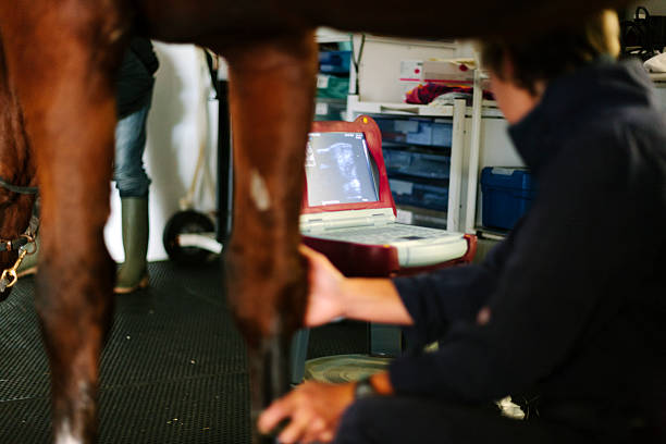
A veterinary ultrasound system generally consists of three main components.
Ultrasound unit
The main unit is responsible for:
- Processing signals received from the transducer.
- Generating and displaying images.
- Adjusting imaging parameters.
- Saving images or video where supported.
- Managing examination information on some models.
Transducer
The transducer both emits and receives ultrasound waves.
Depending on the animal species and intended application, common types may include:
- Linear transducers.
- Convex transducers.
- Microconvex transducers.
- Specialised rectal transducers.
- High-frequency transducers for small animals.
There is no single transducer that is optimal for every species or application.
Software and imaging modes
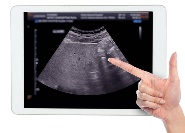
B-mode is the most commonly used mode in reproductive ultrasound.
It provides real-time two-dimensional imaging of structures such as:
- Uterus.
- Gestational sac.
- Embryo or foetus.
- Foetal heartbeat.
- Ovarian follicles.
- Corpus luteum.
- Uterine fluid.
Some systems also include colour Doppler or pulsed-wave Doppler to provide additional information about blood flow.
The presence of Doppler does not automatically mean that B-mode image quality will be better. Image quality also depends on the transducer, processor, system configuration, and operator skill.
1.2. How ultrasound images are produced
The transducer sends sound waves into the animal’s body.
As the waves pass through tissues with different acoustic properties, part of the energy is reflected back to the transducer.
The ultrasound system analyses:
- Time taken for the signal to return.
- Strength of the reflected signal.
- Location of the signal.
and converts this information into an image on the screen.
On an ultrasound image:
- Fluid usually appears black or very dark.
- Soft tissue appears in different shades of grey.
- Strongly reflective structures appear brighter.
Correct interpretation depends heavily on anatomical knowledge and transducer orientation.
1.3. Depth and resolution
Transducer frequency directly affects imaging performance.
Higher frequency
- Better resolution.
- More suitable for superficial structures or small animals.
- Lower penetration depth.
Lower frequency
- Greater penetration.
- More suitable for deeper structures or larger animals.
- Usually lower spatial resolution.
Frequency should therefore be selected according to the depth of the structure being examined rather than simply choosing the sharpest possible image.
2. Main applications of ultrasound in reproductive management
2.1. Pregnancy detection
This is one of the most common uses of reproductive ultrasound.
Ultrasound may help determine:
- Whether an animal is pregnant.
- Presence of a gestational sac or embryo.
- Foetal viability.
- Whether pregnancy appears to be progressing appropriately.
Early identification of non-pregnant animals can allow the farm to:
- Reassess reproductive status.
- Plan rebreeding.
- Reduce unnecessary non-productive waiting periods.
2.2. Evaluation of the uterus and ovaries
Ultrasound may support examination of:
- Ovarian follicles.
- Corpus luteum.
- Abnormal cystic structures.
- Uterine fluid.
- Certain reproductive abnormalities.
These findings may be combined with:
- Oestrus records.
- Breeding dates.
- Clinical examination.
- Reproductive history.
to support breeding or treatment planning.
2.3. Estimation of gestational age
Gestational age may be estimated using features such as:
- Gestational sac size.
- Embryonic size.
- Crown–rump length.
- Development of specific anatomical structures.
Accuracy depends on:
- Species.
- Stage of pregnancy.
- Structure being measured.
- Operator experience.
Gestational-age information may support:
- Estimation of expected parturition date.
- Dry-off planning.
- Nutritional management.
- Preparation of calving or farrowing areas.
2.4. Assessment of foetal viability
Ultrasound may help assess foetal viability through:
- Foetal heart activity.
- Foetal movement.
- Development appropriate for gestational age.
- Other relevant imaging findings.
Foetal death should not be concluded from a single isolated image without appropriate assessment of the relevant signs.
2.5. Determining litter size
In species such as sheep and goats, ultrasound may help distinguish:
- Singleton pregnancy.
- Twin pregnancy.
- Multiple pregnancy.
This information can be valuable for nutritional management, particularly during late pregnancy.
However, counting accuracy depends strongly on timing. When foetuses become larger and overlap, accurate counting may become more difficult.
3. Reproductive ultrasound in cattle
3.1. Timing of pregnancy diagnosis
In cattle, pregnancy can sometimes be detected relatively early by experienced operators using appropriate equipment.
For practical herd management, approximately 28–32 days after breeding is commonly used as an early window for pregnancy confirmation with better practical reliability than very early scanning.
Results still depend on:
- Equipment quality.
- Transducer type and frequency.
- Scanning technique.
- Operator experience.
- Individual-animal factors.
If scanning is performed very early and the result is negative, a repeat examination may be required before concluding that the cow is not pregnant.
3.2. Pregnancy rechecks
Some reproductive programmes include an additional examination after the first pregnancy confirmation.
Objectives may include:
- Confirming continued pregnancy development.
- Detecting early pregnancy loss.
- Updating gestational age.
- Adjusting management groups.
Recheck frequency should be based on herd size and reproductive-management strategy rather than applying one fixed schedule to every farm.
3.3. Ovarian evaluation
In addition to pregnancy confirmation, transrectal ultrasound may help visualise:
- Follicles.
- Corpus luteum.
- Ovarian activity.
- Certain abnormal structures.
This information can be particularly useful in cows that are not pregnant after breeding.
However, placing a cow into an oestrus-synchronisation programme or administering reproductive hormones should not be based only on a finding of “not pregnant.”
Additional factors should be considered, including:
- Ovarian status.
- Presence or absence of a corpus luteum.
- Stage of the reproductive cycle.
- Reproductive history.
- Herd reproductive protocol.
3.4. Applying ultrasound findings to herd management
Pregnant cows may be grouped according to:
- Stage of pregnancy.
- Expected calving date.
- Nutritional status.
- Dry-off schedule.
Non-pregnant cows may undergo:
- Repeat ovarian evaluation.
- Oestrus monitoring.
- Rebreeding.
- Assessment for reproductive disorders.
Animals with repeated breeding failure or recurrent pregnancy loss may be placed under closer reproductive monitoring.
Culling decisions should not be based on a single ultrasound result alone.
They should also consider:
- Age.
- Productivity.
- Reproductive history.
- Medical history.
- Genetic value.
- Economic value.
4. Applications in sows, sheep, goats,
4.1. Sows
In sows, ultrasound is commonly performed transabdominally.
Pregnancy may be detected from around day 20 after breeding, but reliability generally improves noticeably from approximately day 23–24 onward under suitable examination conditions.
Scanning too early may increase the risk of:
- False-negative results.
- Difficulty identifying gestational structures.
- Misinterpretation of fluid-filled structures.
If an early scan is negative but the sow has not returned to oestrus, repeat scanning may be preferable to immediately concluding that she is not pregnant.
Ultrasound may also support evaluation of:
- Ovarian activity.
- Follicles.
- Corpus luteum.
However, using these findings to determine hormone treatment or rebreeding should involve appropriate professional assessment.
4.2. Sheep and goats
Transabdominal ultrasound is commonly used to:
- Confirm pregnancy.
- Estimate gestational age.
- Monitor pregnancy development.
- Determine foetal number.
In sheep, approximately 45–50 days of gestation is often a useful period for litter-size assessment.
As pregnancy advances, overlapping foetal structures may make counting more difficult.
In goats, foetal counting also depends strongly on examination timing and operator experience.
Information on litter size can help support:
- Nutritional adjustment.
- Grouping.
- Late-pregnancy monitoring.
- Preparation for parturition.
5. Ultrasound examination procedure and technique
5.1. Preparing the animal
Before scanning:
- Confirm the correct animal.
- Review breeding records.
- Restrain the animal safely.
- Minimise stress.
- Arrange a suitable working position.
Restraint methods differ between:
- Cattle.
- Pigs.
- Sheep.
- Goats.
- Small animals.
5.2. Preparing the equipment
Check:
- Battery or power source.
- Transducer.
- Transducer cable.
- Ultrasound gel.
- System settings.
- Storage or data-saving functions if required.
An appropriate preset can reduce adjustment time under farm conditions.
5.3. Preparing the operator
The operator should understand:
- Reproductive anatomy.
- Image orientation.
- Transducer handling.
- Normal anatomical structures.
- Structures that may cause confusion.
Practical skill has a major influence on diagnostic accuracy.
5.4. Transrectal ultrasound in cattle
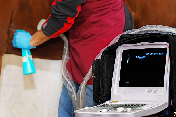
In cattle, a transrectal linear transducer is commonly used to examine the uterus and ovaries.
The operator should:
- Wear appropriate protective gloves.
- Use suitable lubricant.
- Handle tissues gently.
- Avoid excessive force.
- Restrain the cow safely.
- Avoid excessive bending or pulling of the transducer.
Transrectal ultrasound should be performed by someone who has received practical training.
Forceful or incorrect technique may cause:
- Rectal injury.
- Bleeding.
- Harm to the animal.
- Injury risk to the operator.
5.5. Transabdominal ultrasound
This method is commonly used in:
- Pigs.
- Sheep.
- Goats.
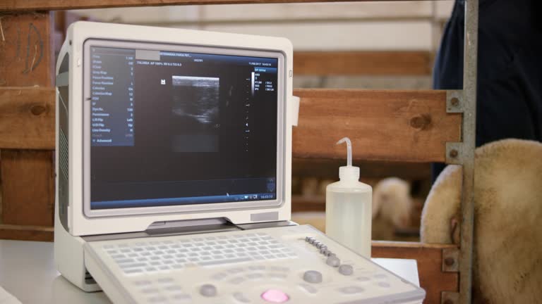
The transducer is placed against the abdominal wall using ultrasound gel.
In pigs, the examination area is often around the:
- Inguinal region.
- Lower abdomen.
In sheep and goats, scanning position changes with gestational stage and technique.
5.6. Role of ultrasound gel
Ultrasound gel removes the air layer between the transducer and the skin.
This helps:
- Improve sound transmission.
- Reduce artefacts.
- Improve image quality.
Insufficient gel or poor transducer contact may produce dark, incomplete, or difficult-to-interpret images.
6. Safety and common errors
6.1. Ultrasound safety
Diagnostic B-mode ultrasound generally has a good safety profile when used appropriately.
However, a suitable principle is ALARA – using the lowest acoustic output and shortest scanning time reasonably necessary to achieve the diagnostic objective.
Operators should:
- Avoid unnecessarily prolonged scanning.
- Use appropriate settings.
- Avoid holding the transducer in one position for excessive periods.
- Avoid excessive mechanical pressure.
- Minimise stress to the animal.
Doppler, particularly pulsed-wave Doppler, generally uses higher acoustic output than B-mode and should therefore be used more cautiously in foetal assessment.
6.2. Common technical errors
Common mistakes include:
- Insufficient gel.
- Incorrect transducer placement.
- Incorrect scanning angle.
- Inappropriate depth settings.
- Gain set too high or too low.
- Scanning too early.
- Reaching conclusions too quickly.
- Missing relevant structures.
- Failing to scan the full required area.
- Failing to compare findings with breeding records.
- Failing to record results.
In cattle, other errors may include:
- Mistaking the urinary bladder for a fluid-filled reproductive structure.
- Misidentifying follicles and the corpus luteum.
- Incorrectly estimating gestational age.
6.3. Transducer hygiene
The transducer should be cleaned appropriately between examinations.
The objectives are to:
- Reduce the risk of pathogen transmission.
- Protect the transducer surface.
- Maintain image quality.
Cleaning agents that may damage transducer materials should not be used unless permitted by the manufacturer.
7. Types of veterinary ultrasound systems
7.1. Portable grayscale ultrasound systems
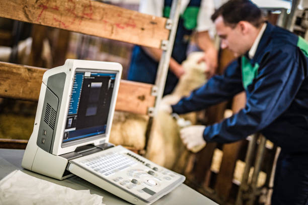
Advantages:
- Compact.
- Easy to carry.
- Suitable for barn or field use.
- Often lower initial investment cost.
- May be sufficient for basic pregnancy diagnosis.
Limitations:
- Smaller display.
- Fewer advanced functions.
- Storage and connectivity capabilities may be limited depending on the model.
Portable systems should not automatically be assumed to produce lower image quality than larger systems; actual performance depends on the model, transducer, and image-processing technology.
7.2. Colour Doppler ultrasound systems
Colour Doppler adds the ability to assess:
- Direction of blood flow.
- Presence of blood flow.
- Certain perfusion characteristics.
Potential applications include:
- Advanced reproductive evaluation.
- Assessment of selected vascular structures.
- Research or specialised diagnostics.
Colour Doppler does not automatically make B-mode images sharper.
Common limitations include:
- Higher cost.
- Greater operator-skill requirements.
- Need for appropriate Doppler use in foetal assessment.
7.3. Cart-based ultrasound systems
Cart-based systems often provide:
- Larger displays.
- Multiple transducer ports.
- Greater expandability.
- More imaging modes.
- Better data-storage capability on many models.
Limitations include:
- Lower mobility in barns or field conditions.
- Greater need for suitable power and working space.
These systems are generally better suited to veterinary clinics, hospitals, or specialist centres.
7.4. Comparison table
| System type | Advantages | Limitations | Suitable applications |
| Portable grayscale | Compact, flexible, easy to use in barns | Screen size and features depend on model | On-farm pregnancy checks in cattle, pigs, sheep, and goats |
| Colour Doppler | Adds information on blood flow and perfusion | Higher cost and greater skill requirements | Advanced reproductive diagnostics, veterinary clinics |
| Cart-based system | Large screen, multiple probes, more functions | Less convenient for field movement | Clinics, hospitals, veterinary centres |
8. Criteria for selecting a veterinary ultrasound system
8.1. Animal species and transducer type
The primary animal group should be identified first.
For example:
- Cattle: often require a linear rectal transducer.
- Pigs: commonly use transabdominal transducers.
- Sheep and goats: require transducers suitable for abdominal depth.
It is better to choose the intended transducer and application first rather than purchasing the machine and then trying to find a suitable probe afterward.
8.2. Frequency range
A suitable frequency range and transducer combination help balance:
- Resolution.
- Penetration depth.
Mixed-species farms may require systems that support multiple transducers or a wider range of frequencies.
8.3. Portability
For beef farms or grazing systems, useful features may include:
- Low weight.
- Good battery life.
- Outdoor-readable display.
- Suitable resistance to dust and moisture.
- Shoulder straps or designs that are easy to carry.
8.4. Durability and battery performance
Farm environments may expose equipment to:
- Dust.
- Moisture.
- Impacts.
- Temperature fluctuations.
Practical durability may therefore be more important than having many advanced functions that are rarely used.
8.5. Data storage and export
For farms seeking data-driven reproductive management, useful capabilities may include:
- Image storage.
- Video storage.
- Individual animal ID.
- Data export.
- Connection to software or computers.
8.6. Maintenance and technical support
Purchase price is not the only cost to consider.
Other considerations include:
- Replacement transducer costs.
- Batteries.
- Warranty.
- Repair turnaround time.
- Technical support.
- Spare-part availability.
A low-cost machine that is difficult to repair or lacks replacement transducers may create higher long-term costs.
8.7. Operator skill level
More functions do not always make a machine more suitable.
New users may benefit from:
- Simple interface.
- Clear presets.
- Fewer operational steps.
- Stable image quality.
In contrast, veterinarians performing advanced diagnostics may require:
- Doppler.
- Multiple transducers.
- Advanced measurements.
- Better data-management functions.
9. Integrating ultrasound data into herd management
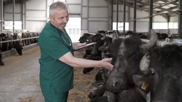
The value of an ultrasound system extends beyond confirming whether an individual animal is pregnant.
When information is recorded at the individual-animal level, farms can build a more continuous reproductive-management system.
9.1. Data that may be recorded
Relevant data may include:
- Animal ID.
- Breeding date.
- Ultrasound examination date.
- Pregnancy status.
- Estimated gestational age.
- Expected parturition date.
- Uterine status.
- Ovarian status.
- Foetal number where assessable.
- Recheck schedule.
9.2. Supporting breeding decisions
If non-pregnant animals are identified early, farms may:
- Monitor oestrus.
- Evaluate ovarian status.
- Schedule rebreeding.
- Enrol animals in an appropriate reproductive programme.
Hormonal treatment or synchronisation protocols should also consider ovarian findings and herd reproductive protocols rather than relying solely on a “not pregnant” result.
9.3. Herd grouping
Results may support grouping animals as:
- Pregnant.
- Non-pregnant.
- Rebreeding required.
- Approaching parturition.
- At risk of pregnancy loss.
- Showing reproductive abnormalities.
- Requiring closer monitoring.
This can support:
- Nutrition.
- Dry-off scheduling.
- Preparation of maternity areas.
- Health monitoring.
9.4. Supporting culling decisions
Animals with:
- Repeated breeding failure.
- Recurrent pregnancy loss.
- Persistent reproductive disorders.
- Poor reproductive performance.
may be flagged for further evaluation.
However, culling decisions should also consider:
- Age.
- Productivity.
- Medical history.
- Genetic value.
- Ongoing care or production costs.
- Potential for reproductive recovery.
9.5. Reducing the cost of non-productive animals
Non-pregnant animals still consume:
- Feed.
- Labour.
- Housing space.
- Health-management resources.
Earlier detection helps farms make more timely decisions about:
- Rebreeding.
- Treatment.
- Monitoring.
- Culling.
rather than continuing to maintain animals without a defined reproductive plan.
10. Checklist for reproductive ultrasound on farms
Before scanning
- Review breeding records.
- Confirm animal identity.
- Define the examination objective.
- Check battery or power supply.
- Check transducer condition.
- Prepare ultrasound gel.
- Prepare gloves and lubricant where required.
- Select the appropriate preset.
- Restrain the animal safely.
During scanning
- Position the transducer correctly.
- Adjust imaging depth appropriately.
- Set gain correctly.
- Scan the full required area.
- Avoid reaching conclusions too early.
- Evaluate the uterus, pregnancy, and ovaries where relevant.
- Use Doppler only where necessary.
- Follow the ALARA principle.
- Record or save findings.
After scanning
- Clean the transducer according to instructions.
- Record the examination date.
- Update reproductive status.
- Save images where useful.
- Schedule rechecks.
- Schedule rebreeding where appropriate.
- Move animals between management groups where required.
- Synchronise findings with the farm-management system where available.
11. Frequently asked questions about veterinary ultrasound
What is veterinary ultrasound used for?
In reproductive management, ultrasound may be used to:
- Confirm pregnancy.
- Evaluate the uterus.
- Evaluate the ovaries.
- Visualise follicles and corpus luteum.
- Estimate gestational age.
- Assess foetal viability.
- Determine foetal number in some species.
- Monitor certain reproductive abnormalities.
Outside reproduction, ultrasound is also widely used for soft-tissue and abdominal imaging.
How long after breeding should cattle be scanned for pregnancy?
Approximately 28–32 days after breeding is a commonly used early window in herd management.
Pregnancy may sometimes be detected earlier under suitable conditions, but very early examination increases the risk of false-negative results.
If an early scan is negative, repeat examination may be required.
When is pregnancy ultrasound in sows considered more reliable?
Pregnancy may be detected from around day 20.
In practice, reliability generally improves from approximately day 23–24 onward.
If an early scan is negative but the sow has not returned to oestrus, repeat examination is recommended.
Can ultrasound accurately count foetuses?
This depends on the species and timing.
In sheep and goats, ultrasound may help determine foetal number when performed at an appropriate stage.
In sheep, approximately 45–50 days is often a useful period for foetal counting.
Can repeated ultrasound examinations affect the foetus?
Diagnostic B-mode ultrasound generally has a good safety profile when used appropriately.
However, operators should still:
- Avoid unnecessary prolonged scanning.
- Use appropriate acoustic output.
- Follow the ALARA principle.
- Use Doppler more cautiously during foetal assessment.
How much training does an operator need?
Basic pregnancy detection can often be learned relatively quickly with:
- Proper instruction.
- Anatomical knowledge.
- Sufficient supervised practice.
More advanced applications such as:
- Ovarian assessment.
- Differentiation of follicles and corpus luteum.
- Accurate gestational-age estimation.
- Diagnosis of reproductive disease.
- Doppler imaging.
require considerably more experience.
When should transrectal palpation be combined with ultrasound?
In cattle, rectal palpation and ultrasound can complement each other.
Manual palpation provides information about:
- Size.
- Consistency.
- Position of structures.
Ultrasound provides more direct imaging information about:
- Fluid.
- Follicles.
- Corpus luteum.
- Embryo or foetus.
Using both methods may improve diagnostic confidence in certain situations.
Can progesterone testing replace ultrasound?
No.
Progesterone testing may help assess:
- Corpus luteum activity.
- Reproductive cycle.
- Certain situations where additional data are useful.
However, interpretation depends on species and sampling time.
For example, a high progesterone concentration does not always confirm pregnancy.
Ultrasound, clinical examination, and hormone testing should therefore be regarded as complementary tools where appropriate.
Using Ultrasound to Improve Herd Reproductive Efficiency at VIETSTOCK 2026
Veterinary ultrasound provides greater value than simply confirming whether an animal is pregnant. When ultrasound findings are combined with breeding dates, ovarian status, gestational age, and individual-animal data, farms can make more informed decisions about breeding, herd grouping, preparation for parturition, and reproductive performance.
At VIETSTOCK 2026 – Vietnam’s Premier International Feed, Livestock & Meat Industry Show, farmers, veterinarians, and businesses can explore new developments in livestock production, veterinary medicine, and farm technology while connecting directly with organisations across the industry value chain. The event is expected to bring together 300 brands, over 10,000 sqm of exhibition area, and 13,000 trade visitors from 40 countries.
For farms seeking to improve reproductive performance and make better use of herd data, participants can explore:
- Veterinary equipment and technology: exploring solutions for diagnosis, health monitoring, and reproductive assessment.
- Breeding and reproductive solutions: learning about approaches that support breeding, pregnancy monitoring, and improved reproductive efficiency.
- Farm data management: exploring tools for storing individual-animal information, breeding records, examination results, and expected parturition dates to support decision-making.
- Herd-monitoring technology: exploring solutions that connect health, reproductive, and productivity data during farm operations.
- Stage-specific nutrition and care: learning about solutions for pregnant animals, preparation for parturition, and postpartum recovery.
For participants interested in using data to improve livestock-production efficiency, the One Health Forum on 22 October 2026 also provides a relevant perspective. The programme includes topics such as “Integrating One Health into commercial livestock production systems” and “AI and data analytics in herd/flock One Health management.” These discussions provide a useful broader context for how ultrasound findings, reproductive records, and other herd-management data can contribute to farm decision-making.
Date: 21–23 October 2026
Venue: Saigon Exhibition and Convention Center (SECC), 799 Nguyen Van Linh Street, Tan My Ward, Ho Chi Minh City, Vietnam
Event website: https://www.vietstock.org/en/
Register to visit now: https://ers-vn.informa-info.com/vs26
👉 VIETSTOCK 2026 Visitor Registration Information
👉 Learn more about the One Health Forum
Contact information:
Exhibiting: Ms. Sophie Nguyen – [email protected]
Visitor Support: Ms. Phuong – [email protected]
Marcom Support: Ms. Anita Pham – [email protected]