Veterinary Clinical Diagnosis: Examination Process, Clinical Sign Assessment, and When Testing Is Needed
Veterinary clinical diagnosis helps veterinarians identify health abnormalities, assess clinical signs, develop preliminary diagnoses, and determine when additional diagnostic testing is needed for effective herd and flock health management.
Veterinary clinical diagnosis is a foundational first step in identifying diseases in animals. Through observation, history taking, and direct examination, veterinarians can develop an initial diagnostic hypothesis, assess disease severity, and decide whether additional ancillary diagnostic testing is needed. Farm technicians can support the process by collecting data, monitoring clinical signs, and providing herd or flock history under appropriate professional guidance.
A systematic approach to veterinary disease diagnosis helps improve the quality of information used for professional decision-making. It also reduces the risk of testing or using medicines based on assumptions and helps farms respond more promptly to abnormalities in the herd or flock. However, when serious infectious diseases are suspected, mortality increases rapidly, clinical signs are atypical, or animals do not respond to the initial management approach, veterinary consultation and appropriate testing are needed.
Key takeaways
- Veterinary clinical diagnosis is a systematic process that includes history taking, observation, organ-system examination, and data synthesis to develop a preliminary diagnosis.
- Clinical diagnosis and ancillary diagnostics complement each other: clinical findings guide disease suspicion, while ancillary diagnostics help confirm or clarify unresolved points.
- Clinical signs should be classified by specificity, organ system, and disease progression to avoid misinterpretation or drawing conclusions from a single sign.
- Testing should be considered when clinical findings are not sufficient, disease progression is unusual, or a serious infectious disease is suspected.
- Common mistakes include overlooking herd or flock history, examining only individual animals, and failing to record data systematically. Veterinary medicines or antibiotics should not be changed without an adequate diagnostic basis.
What Is Veterinary Clinical Diagnosis and Why Does It Matter in Veterinary Practice?
Definition and Basic Terms
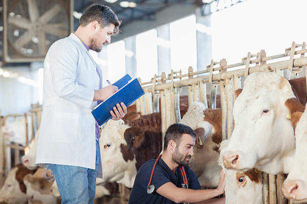
Veterinary clinical diagnosis is the process of collecting and analyzing information from case history, direct observation, and physical examination of animals to assess disease status, before or alongside the use of ancillary diagnostic methods when needed. It is a core skill in veterinary practice and requires a combination of knowledge in anatomy, physiology, pathology, epidemiology, and field experience.
Key terms to distinguish include:
- Clinical signs: observable or examination-based signs of disease, such as fever, coughing, diarrhea, or loss of appetite.
- Syndrome: a group of clinical signs that often occur together and may suggest a certain group of diseases or causes.
- Preliminary diagnosis: a disease hypothesis developed based on clinical and epidemiological data, but not yet fully confirmed.
- Definitive diagnosis: a conclusion reached after sufficient clinical, epidemiological, and/or ancillary diagnostic evidence has been reviewed.
Clinical, Ancillary, Preliminary, and Definitive Diagnosis
Clinical diagnosis is mainly based on information that veterinarians collect through observation, history taking, palpation, auscultation, percussion, measurement of physiological parameters, and direct examination of animals. Ancillary diagnostics add data from methods such as blood tests, fecal analysis, PCR, ELISA, diagnostic imaging, microbial culture, histopathology, or necropsy.
These two approaches are not separate or competing processes. They complement each other. Clinical findings help guide the suspected disease group and the selection of appropriate diagnostic tests and testing objectives. Ancillary diagnostics help confirm the causative agent, assess lesions, or rule out causes with similar clinical presentations.
In some cases where the disease pattern is familiar, clinical signs are relatively typical, and there are no warning signs of a serious infectious disease, veterinarians may consider an initial management approach based on clinical assessment. The use of veterinary medicines should still follow the product label, professional guidance, and current regulations. For complex cases, unusual progression, or diseases that require control measures, appropriate testing should be combined with clinical assessment.
Role in Herd Health Management and Treatment Decisions
In intensive livestock production, veterinary clinical diagnosis is not limited to individual animals. It is also an important tool for herd or flock health surveillance. Early detection of changes in feed intake, behavior, respiratory signs, digestive signs, mortality, or productivity can help farms identify abnormalities before problems spread.
Clinical information also provides a basis for veterinarians to consider isolating suspected animals, increasing monitoring, evaluating disease prevention programs, selecting diagnostic tests, or recommending an appropriate management approach. When a notifiable or controlled infectious disease is suspected, the relevant local veterinary authority should be contacted in accordance with current regulations.
Objectives and Basic Principles of Veterinary Clinical Diagnosis
Objectives: Disease Detection, Treatment Direction, and Appropriate Testing
The direct objectives of clinical diagnosis include:
- Identifying whether animals show abnormal signs and assessing disease severity.
- Guiding the likely disease group or cause group so veterinarians can consider appropriate monitoring, testing, or management.
- Deciding whether ancillary diagnostic testing is needed and, if so, selecting the test group that matches the diagnostic objective.
- Assessing prognosis, risk of spread, and intervention priority within the herd or flock.
Professional Principles: Objective, Systematic, and Evidence-Based
Effective clinical diagnosis should follow several core principles:
- Objectivity: avoid allowing assumptions or experience from previous cases to dominate the conclusion. Findings should be recorded as observed before they are interpreted.
- Systematic process: examine animals in a consistent sequence to reduce the risk of missing important signs, while adjusting priorities according to the actual case.
- Evidence-based reasoning: combine history, herd or flock data, epidemiological information, and physical signs. Do not conclude based on a single sign alone.
Biosecurity and Animal Welfare Principles During Examination
When conducting clinical examinations on farms, it is important to ensure:
- Appropriate personal protective equipment, especially when an infectious disease or zoonotic risk is suspected.
- Limited unnecessary movement between housing areas to reduce the risk of cross-contamination.
- An examination order that moves from lower-risk areas to suspected disease areas when practical conditions allow.
- Reduced stress, injury, and restraint time during examination, especially for poultry, piglets, or weakened animals.
Step-by-Step Veterinary Clinical Examination Process
Preparation Before Examination: Tools, Personnel, and Housing Conditions
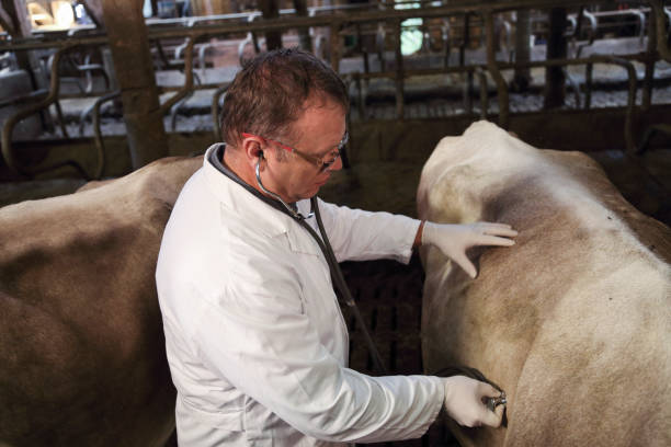
Before starting the examination, prepare:
- Basic tools: thermometer, stethoscope, flashlight, gloves, personal protective equipment, cleaning and disinfection tools, and a notebook or digital device for recording data.
- Support personnel: someone experienced in handling animals and someone who understands the herd or flock management history.
- Housing conditions: adequate lighting for observation, reduced noise to limit stress, and a plan to separate suspected animals when needed.
Step 1 – History Taking for Individual Animals and the Herd or Flock
History taking is important but is often done too briefly. Information to collect includes:
- Species, breed, age, weight, sex, and number of affected animals.
- Time of onset of the first clinical signs and their progression over time.
- Morbidity rate, mortality rate, and changes in these indicators within the herd or flock.
- Vaccination history, parasite control, previous treatments, and medicines used.
- Diet, water source, and recent feed changes.
- Recent introduction of new animals, contact with other herds or flocks, or changes in housing conditions.
- Similar diseases previously seen on the farm or the epidemiological situation in the area.
If animals were medicated before examination, record the product name, dosage, route of administration, duration of use, and most recent time of administration. These details can significantly affect clinical presentation and interpretation of test results.
Herd or flock history often provides essential context for interpreting clinical signs observed in an individual animal.
Step 2 – General Observation: Posture, Behavior, Nutritional Status, Mucous Membranes, and Feces
Before approaching the animals, observe from a distance and record:
- Posture and behavior: standing, lying down, separating from the group, abnormal movement, seizures, or loss of balance.
- Nutritional status: body condition, skin, hair or feathers, and uniformity within the herd or flock.
- Mucous membranes: the color of the ocular, nasal, and oral mucous membranes, such as pale, yellow, bluish, or other abnormal changes.
- Feces and urine: color, consistency, frequency, and the presence of blood or mucus if observed.
- Respiration: respiratory rate, breathing pattern, open-mouth breathing, and abnormal sounds.
General observation of the herd or flock before individual examination helps assess how widespread the problem is and supports the selection of representative animals at different stages of clinical presentation.
Step 3 – Detailed Examination by Organ System
After general observation, conduct a systematic examination by organ system. Key systems to assess include:
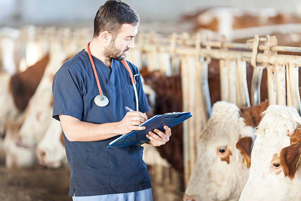
- Respiratory system: auscultate the lungs when appropriate, check nasal discharge, and evaluate respiratory rate and breathing pattern.
- Digestive system: assess digestive function, palpate the abdomen when appropriate, examine the oral mucosa, and assess fecal condition.
- Circulatory system: measure body temperature and evaluate heart rate, pulse, and perfusion when needed.
- Reproductive and urinary systems: check for abnormal discharge, udder condition, reproductive organs, and urine depending on the species and clinical situation.
- Nervous and musculoskeletal systems: assess gait, reflexes, muscle tone, ability to stand, and abnormalities in joints or limbs.
The examination should not stop as soon as an abnormality is found in one organ system. Some diseases may show prominent signs in one system while also affecting others. The detailed examination process should be adjusted according to species, age, animal condition, and the actual disease presentation.
Step 4 – Recording and Synthesizing Examination Data
After completing the examination, record:
- Identification information for the individual animal and the herd or flock.
- Observed clinical signs, including negative signs that are diagnostically useful, such as no fever or no nasal discharge.
- Measured results such as body temperature, heart rate, and respiratory rate when measured.
- Preliminary assessment and a list of differential diagnoses.
- Next decision: continued monitoring, isolation, sample collection for testing, or management/treatment according to appropriate professional guidance.
Systematic record keeping is essential for tracking disease progression, evaluating the response to intervention, and building herd or flock health data over time.
Classification and Assessment of Clinical Signs in Livestock and Poultry
Clinical Sign Classification: Specific and Non-Specific, Primary and Secondary, Acute and Chronic
Not all clinical signs have the same diagnostic value. Proper classification helps reduce misinterpretation:
- Highly suggestive signs: certain signs may increase suspicion of a specific disease group. For example, vesicular lesions in the mouth or on the feet may raise suspicion of foot-and-mouth disease in susceptible species, but a single sign should not be used to confirm a disease on its own.
- Non-specific signs: signs such as fever, loss of appetite, or reduced weight gain can occur in many different diseases. They must be combined with history and epidemiological context to have diagnostic value.
- Primary signs: the most prominent signs, often the reason why the farm reports the problem.
- Secondary signs: accompanying signs that can help narrow the differential diagnosis.
- Acute signs: signs that appear quickly and progress over a short period; these may occur in infectious diseases, poisoning, or many other acute conditions.
- Chronic signs: signs that persist or recur and may be related to parasitic disease, nutrition, metabolic disorders, prolonged infection, or other causes.
Common Clinical Sign Groups by Organ System
Common clinical sign groups in on-farm veterinary diagnosis include:
- Respiratory signs: coughing, sneezing, nasal discharge, rapid breathing, difficulty breathing, or abnormal respiratory sounds. Depending on species, age, and epidemiology, these signs may occur in many respiratory diseases such as pneumonia in pigs, PRRS, Mycoplasma infection, or avian influenza in poultry.
- Digestive signs: diarrhea, feces with blood or mucus, reduced feed intake, bloating, or changes in feces. These signs may occur in many intestinal diseases, coccidiosis, feed-related disorders, poisoning, or infectious diseases depending on the species.
- Nervous signs: seizures, circling, loss of balance, paralysis, or behavioral changes. This sign group may be related to infectious diseases, inflammatory neurologic conditions, nutritional deficiency, poisoning, or metabolic disorders. Further assessment is needed before identifying the cause.
- Skin and mucous membrane signs: hemorrhage, vesicles, ulcers, jaundice, or pale mucous membranes. These signs may suggest multiple disease groups and should be interpreted in the context of species, epidemiology, and accompanying signs.
- Reproductive signs: abortion, stillbirth, reduced conception rate, or abnormal genital or reproductive tract discharge. Causes may include infectious diseases, nutrition, heat stress, toxins, or reproductive management issues.
Factors That May Interfere With Clinical Signs: Prior Medication, Stress, and Feed Changes
Several factors can change or mask clinical signs:
- Medication before examination: antibiotics, antipyretics, anti-inflammatory drugs, or other medicines can change the clinical presentation. Ask clearly about the product name, dosage, route of administration, and time of use.
- Transport stress or environmental change: may cause increased heart rate, rapid breathing, reduced feed intake, or temporary digestive disturbance.
- Sudden feed changes: may cause digestive disorders and should be considered when differentiating infectious from non-infectious causes.
- Physiological stage: pregnancy, post-partum period, lactation, growth, or other production stages may change some normal physiological parameters.
From Clinical Signs to Preliminary Diagnosis and Differential Diagnosis
Developing a Preliminary Diagnostic Hypothesis Based on History, Epidemiology, and Clinical Signs
A preliminary diagnosis is the result of systematic reasoning, not guesswork. The process includes:
- Summarizing the primary and secondary signs recorded.
- Comparing them with herd or flock history, including age, vaccination program, previous treatment, and regional epidemiology.
- Identifying the main affected organ system and any related systems.
- Listing diseases or cause groups that could produce the clinical picture.
- Prioritizing possibilities based on epidemiology, consistency with the clinical signs, disease severity, and the need for urgent intervention.
Building a Differential Diagnosis List for Respiratory, Digestive, and Reproductive Disease Groups
Differential diagnosis is a list of diseases or cause groups that should be considered, ruled out, or confirmed. Examples include:
- Respiratory syndrome in pigs: PRRS, swine influenza, Mycoplasma hyopneumoniae, Actinobacillus pleuropneumoniae (APP), Pasteurella multocida, and other agents may be considered depending on the case. Age group, morbidity and mortality rates, accompanying clinical signs, vaccination program, and epidemiological situation should be assessed.
- Diarrhea in poultry: may be related to coccidiosis, necrotic enteritis, salmonellosis, feed changes, or other infectious diseases. The cause should not be concluded based only on fecal color. Age, mortality rate, flock-level signs, vaccination program, lesions, and testing should be considered when needed.
- Reproductive disorders: may be related to PRRS in pigs, brucellosis, leptospirosis, nutrition, toxins, heat stress, or many other causes. Reproductive history, epidemiology, and appropriate tests for suspected agents should be combined.
Detailed differential diagnosis guidance for each disease group is covered in specialized articles within this content series.
When Empirical Treatment May Be Considered and When Caution Is Needed
In some low-risk situations, veterinarians may consider an initial management approach based on clinical experience when:
- The disease presentation is familiar, clinical signs are relatively typical, and the case has been assessed in the farm’s epidemiological context.
- Morbidity is low, with no rapid increase in mortality or unusual spread.
- The response can be monitored over a period appropriate to the case, type of intervention, and professional guidance.
The use of veterinary medicines, especially antibiotics, should be based on professional assessment, product label directions, current regulations, and withdrawal period requirements when applicable. Medicines should not be changed or combined without a clear diagnostic basis.
Caution and testing should be prioritized when:
- Mortality is high or increases unusually quickly.
- Animals do not respond to the initial management approach within a timeframe appropriate to the case.
- A serious infectious disease subject to mandatory notification, outbreak declaration, or control under current regulations is suspected.
- Clinical signs are atypical, multiple diseases have similar presentations, or clinical data are insufficient to narrow the diagnosis.
When Should Diagnostic Testing Be Ordered in Veterinary Disease Diagnosis?
Situations Where Testing Should Be Prioritized
In some situations, clinical diagnosis alone may not be sufficient and appropriate testing should be considered, including:
- Suspected serious infectious diseases that are subject to mandatory notification, outbreak declaration, or control measures under regulations: the relevant authority should be contacted and sample collection/testing should follow applicable guidance.
- High mortality or rapid progression that cannot be explained by clinical data: veterinarians may consider necropsy, sample collection, and tests that match the clinical picture.
- No response to initial treatment: the diagnosis and medication history should be reassessed. Identifying the causative agent or performing antimicrobial susceptibility testing may be considered when a bacterial cause is suspected and testing is professionally indicated.
- Need to assess organ function: hematology or blood biochemistry may be used depending on species, case presentation, and diagnostic objective.
- Herd or flock health and immune status monitoring: surveillance programs may use serological tests or other methods depending on the disease, vaccine, and management objective.
When warning signs are observed, such as a sudden increase in mortality, abnormal hemorrhage, widespread neurological signs in the herd or flock, or suspicion of a zoonotic disease, early professional assessment should be prioritized. The competent veterinary authority should be contacted when the case falls under reporting requirements.
When Clinical Diagnosis May Be Sufficient for a Treatment Decision
In some cases, veterinarians may recommend an initial management approach based on clinical data when:
- The disease presentation is relatively typical, consistent with the farm’s history and epidemiology, and there are no warning signs of a serious infectious disease.
- Morbidity is low, the herd or flock remains stable, and disease progression can be closely monitored after intervention.
- The case does not fall into a group that requires confirmatory testing or a specific control protocol under current regulations.
Whether testing is needed should be weighed against the information value of the test, disease risk, cost, and practical conditions. Testing should not be skipped solely because of cost if the result may significantly change professional decisions or disease control measures.
Suggested Test Groups by Diagnostic Objective
Test selection should be based on the suspected agent, disease stage, sample type, and diagnostic objective. The table below provides general guidance only. Specific sample types, sampling time, and methods should follow the guidance of the veterinarian or receiving laboratory.
| Diagnostic objective | Suitable test group |
| Identify bacterial agents | Bacterial culture and identification; antimicrobial susceptibility testing when indicated |
| Identify viral agents | PCR/RT-PCR, antigen tests, serology, or other methods depending on the agent |
| Assess herd or flock immunity | Antibody tests such as ELISA, HI, or SNT depending on the disease and objective |
| Assess organ function | Blood biochemistry using relevant indicators according to species and case presentation |
| Assess hematologic status | Complete blood count (CBC) and related parameters |
| Identify parasites | Fecal analysis, direct microscopy, or methods suitable for the suspected parasite |
| Assess tissue lesions | Necropsy combined with histopathology sample collection when indicated |
Detailed guidance on sample collection, preservation, submission, and specific testing methods is covered in specialized articles within this content series.
Overview of Veterinary Disease Diagnostic Methods
Clinical Methods
Clinical methods include techniques that collect information directly from animals and the herd or flock, such as observation, history taking, palpation, auscultation, percussion, and measurement of basic physiological parameters. These methods can be performed on the farm using basic examination tools and help guide the next diagnostic steps.
Ancillary Methods: Laboratory Testing, Diagnostic Imaging, and Necropsy
- Laboratory testing: may include microbiology, PCR, ELISA, hematology, biochemistry, parasitology, or histopathology depending on the objective. These methods provide additional objective evidence to identify agents, assess physiological or health status, or clarify differential diagnoses.
- Diagnostic imaging: X-ray, ultrasound, and other techniques may support assessment of organs such as the lungs, heart, abdomen, or reproductive system depending on the species. On-farm application depends on equipment, personnel, cost, and operational conditions at each facility.
- Necropsy: allows direct observation of gross lesions and sample collection for microbiology or histopathology. This method is especially valuable when animals have died or must be culled, but it should be performed by qualified personnel and in compliance with biosecurity requirements.
Epidemiological Methods and Herd/Flock Data Analysis
Analysis of production data such as feed intake, weight gain, feed conversion ratio, mortality rate, laying rate, or reproductive indicators over time can help detect abnormal trends before clinical signs become obvious.
When combined with epidemiological information from the farm and surrounding area, these data support risk identification, selection of animal groups for examination, and prioritization of prevention or early diagnosis measures.
The Place of Clinical Diagnosis in the Overall Diagnostic Process
Clinical diagnosis is an important starting point in veterinary disease diagnosis. It guides the next steps: deciding whether testing is needed, selecting appropriate tests, and interpreting results within the correct clinical and epidemiological context.
A test result should not be separated from case history, clinical signs, sampling time, and herd or flock status. Combining multiple data sources helps reduce the risk of misinterpretation and increases the value of test results in professional decision-making.
Practical Examples: Applying the Clinical Diagnosis Process on Farms
Respiratory Disease Case in a Pig Herd: History, Clinical Examination, Preliminary Diagnosis, and Testing Decision
This is an illustrative example of the process and does not represent a confirmed specific case.
A grow-finish pig farm reports that a significant proportion of pigs aged 8–12 weeks have shown coughing, rapid breathing, reduced feed intake, and fever over 3–4 days. Some pigs have clear nasal discharge. The herd has received PRRS and Mycoplasma vaccines according to the farm’s disease prevention program and applicable professional guidance.
Clinical examination process:
- History taking: no recent introduction of new pigs, no feed change, and a notable weather change during the previous week. The farm has used antibiotics for two days but has not observed clear improvement. The product type, dosage, route of administration, timing, and person who made the treatment decision should be reviewed before assessing response.
- General observation: many pigs separate from the group, lie down more than usual, show abdominal breathing, and some breathe through the mouth.
- Detailed examination: increased body temperature is recorded. Some pigs show abnormal respiratory sounds, clear nasal discharge, and paler-than-normal mucous membranes.
- Preliminary diagnosis: respiratory syndrome requiring differential diagnosis among viral agents, bacterial agents, and co-infections. PRRS cannot be ruled out based only on vaccination history, because vaccine protection depends on multiple factors and vaccination does not replace diagnosis when the herd shows clinical signs.
- Testing decision: because morbidity is high and the herd has not responded to the initial management approach, a veterinarian or laboratory should be consulted to select appropriate sample types and test methods. Depending on the case, respiratory samples, blood/serum, or tissue samples from dead animals may be considered when indicated. If a bacterial cause is suspected, culture and antimicrobial susceptibility testing may support appropriate antibiotic selection.
Lesson learned: lack of response to initial antibiotics is a signal to reassess the diagnosis and medication history. Antibiotics should not be repeatedly changed based only on assumptions when the cause has not been clearly identified.
Digestive Disease Case in a Chicken Flock: Flock Observation, Clinical Sign Assessment, and the Value of Combining Clinical and Ancillary Diagnosis
This is an illustrative example of the process and does not represent a confirmed specific case.
A three-week-old broiler flock has abnormally colored loose droppings, and some birds appear depressed and eat less. Mortality has increased slightly over two days. The flock has not received Newcastle disease vaccination according to the farm’s disease prevention program and is transitioning to phase 2 feed.
Flock observation: abnormal droppings are found in many areas of the house. Some chickens stand with ruffled feathers and closed eyes. No obvious respiratory signs have been observed.
Preliminary assessment: coccidiosis is one cause to consider at this age, but necrotic enteritis, feed change effects, and other infectious diseases should not be ruled out based only on fecal appearance. The lack of Newcastle disease vaccination should be included in the epidemiological assessment, but it is not sufficient to conclude that the flock has Newcastle disease.
Decision: fecal samples or other appropriate samples may be collected to help check for parasites and other intestinal causes. If dead birds are available, a veterinarian may consider necropsy and lesion-based sample collection. When epidemiological factors or clinical signs increase suspicion of Newcastle disease or another serious infectious disease, confirmatory testing should be selected according to professional advice and current regulations.
Lesson learned: in poultry, whole-flock observation and lesion assessment in dead birds can provide important information alongside examination of live birds. However, sample types and test methods should be selected according to the suspected agent and laboratory guidance. A fixed testing procedure should not be applied to every case.
Common Mistakes in Veterinary Clinical Diagnosis and How to Avoid Them
Mistakes in History Taking and Information Collection From Farm Owners
- Overlooking herd or flock history: asking only about the sick individual without considering the overall herd or flock situation, vaccination program, and previous problems.
- Not asking about medicines already used: this can lead to misinterpretation of clinical signs, treatment response, or test results.
- Relying entirely on the farm owner’s description: information may be incomplete or inaccurate. History should be compared with records and actual observation.
Examination Mistakes: Missing Organ Systems and Focusing Only on Individuals Instead of the Herd or Flock
- Examining only the organ system with the most obvious clinical sign: this may miss signs in other organ systems that are valuable for differential diagnosis.
- Examining only one animal: one animal may not represent the whole herd or flock. Multiple animals should be selected according to herd/flock size, disease severity, and different stages of presentation.
- Not observing the herd or flock before approaching individual animals: this can result in loss of information about natural behavior, the distribution of clinical signs, and the level of impact across the group.
Mistakes in Clinical Sign Interpretation and Ignoring Epidemiological Data
- Diagnosing based on a single clinical sign: for example, concluding that fever means infection without evaluating other causes.
- Not updating the local epidemiological situation: this may cause the farm to miss risks related to outbreaks or diseases circulating in the area.
- Ignoring seasonal factors and housing conditions: temperature, humidity, ventilation, stocking density, and weather changes can significantly affect herd or flock health and clinical presentation.
Recommendations to Improve Diagnostic Accuracy: Training, Checklists, and Data Standardization
- Use clinical examination checklists to reduce missed steps, especially for technicians or less experienced veterinarians.
- Conduct regular training on recognition of clinical signs, history taking, and updates on the local epidemiological situation.
- Record examination data and production data consistently to detect abnormal trends.
- Consult experienced veterinarians or laboratories when cases are atypical, do not respond to initial management, or may spread within the herd or flock.
Veterinary Clinical Examination Checklist and Testing Decision Flow

Below is an example of a 10-step veterinary clinical examination checklist for reference. Veterinarians may adjust it according to species and practical conditions.
- Prepare tools and personal protective equipment before entering the animal housing area.
- Take history: individual animal history, herd or flock history, vaccination program, and medicines used.
- Observe the whole herd or flock from a distance before approaching individual animals.
- Select representative animals at different stages of presentation for examination.
- Conduct general observation of the individual animal: posture, behavior, body condition, mucous membranes, and feces.
- Measure parameters such as body temperature, heart rate, and respiratory rate when appropriate for the species and case.
- Examine the respiratory system: assess breathing pattern, nasal discharge, and lung sounds when appropriate.
- Examine the digestive system: assess digestive activity, abdominal area, and fecal condition.
- Examine remaining organ systems based on priorities indicated by the clinical signs.
- Record findings fully, develop a preliminary diagnosis, and decide the next step.
The flow below is a general reference for deciding whether testing is needed. It does not replace guidance from veterinarians, laboratories, or current legal requirements:
Clinical examination completed
↓
Clinical signs are relatively typical + risk is low + no warning signs?
↓ Yes → Manage/monitor according to professional guidance + record data
↓ No
Atypical clinical signs / no response to management / high mortality?
↓ Yes → Reassess diagnosis + select appropriate testing
↓
Suspected serious infectious disease / notifiable disease?
↓ Yes → Contact the competent veterinary authority + collect samples and perform testing according to guidance
↓
Test results + clinical and epidemiological data → Diagnosis → Appropriate management approach
FAQ on Veterinary Clinical Diagnosis
How is veterinary clinical diagnosis different from diagnostic testing?
Clinical diagnosis is mainly based on history, observation, and direct examination using basic examination tools. Testing provides additional evidence about causative agents, immune status, lesions, or biological changes that clinical examination cannot fully determine.
The two methods complement each other. Clinical findings help identify what needs to be clarified and which tests are appropriate. Test results then need to be interpreted in the clinical and epidemiological context of the herd or flock.
Can the same clinical examination process be used for pigs, chickens, and cattle?
The general principles are similar: history taking, herd or flock observation, individual examination, organ-system assessment, record keeping, and preliminary diagnosis. However, examination techniques, normal physiological parameters, restraint methods, and priority disease groups differ significantly between species.
Therefore, the specific process should be adjusted according to species, age, production purpose, and the actual case.
How can two diseases with similar clinical signs be distinguished by clinical examination alone?
When two diseases have similar clinical signs, additional data should be collected, such as age of affected animals, speed of spread, mortality rate, vaccination program, medication history, epidemiological situation, and other signs seen across the herd or flock.
If clinical data are still insufficient to distinguish between diseases, or if the diagnosis may significantly change treatment and disease control decisions, appropriate testing should be selected instead of forcing a conclusion from clinical signs alone.
Can systematic clinical diagnosis help reduce testing costs for farms?
It can. When clinical examination and history taking are conducted systematically, veterinarians can identify the specific question that needs to be answered and select more appropriate tests instead of running multiple tests without a clear objective.
However, the main goal is not to reduce the number of tests at all costs. When testing is important for treatment decisions, disease control, or legal requirements, it should not be skipped simply because of cost.
How should newly graduated veterinarians develop clinical diagnosis skills?
They should prioritize supervised practice with experienced veterinarians and exposure to a wide range of clinical cases. Examination checklists can help build systematic habits. Full case records should be kept, and preliminary diagnoses should be compared with test results or actual case progression to support learning.
In addition to examination skills, veterinarians should regularly update their knowledge of epidemiology, emerging and re-emerging diseases, veterinary regulations, responsible medicine use, and skills in working effectively with diagnostic laboratories.
Connecting Diagnostic Knowledge With Herd Health Management at VIETSTOCK 2026
Veterinary clinical diagnosis is the foundation that helps veterinarians, technicians, and farm owners detect abnormalities early, classify clinical signs, develop preliminary diagnoses, and decide when additional testing is needed. When examination, record keeping, and herd or flock history assessment are conducted systematically, farms gain a stronger basis for timely intervention, reduced assumption-based treatment, and more effective animal health management.
VIETSTOCK 2026 is an industry networking platform for livestock producers, animal health companies, production facilities, technology suppliers, and businesses across the livestock value chain. The event is expected to bring together more than 300 brands, over 10,000 sqm of exhibition area, and 13,000 trade visitors from more than 40 countries and regions.
In relation to diagnosis and disease management, visitors can explore solutions that support herd and flock health monitoring, farm data management, biosecurity, veterinary vaccines, veterinary medicines, nutrition, and technologies that help detect early signs of abnormalities in livestock production.
Alongside the exhibition, VIETSTOCK 2026 will also organise the VIETSTOCK 2026 Livestock Roadshows in Bac Ninh, Dong Thap and Ho Chi Minh City, focusing on sharing practical knowledge on herd and flock management, disease prevention and control, biosecurity, and improving livestock production efficiency in key livestock regions.
Date: 21–23 October 2026
Venue: Saigon Exhibition and Convention Center (SECC), 799 Nguyen Van Linh Street, Tan My Ward, Ho Chi Minh City, Vietnam
Event website: https://www.vietstock.org/en/
👉 Visitor registration for VIETSTOCK 2026
👉 Learn more about the VIETSTOCK 2026 Livestock Roadshows
Contact information:
Exhibiting: Ms. Sophie Nguyen – [email protected]
Visitor Support: Ms. Phuong – [email protected]
Marcom Support: Ms. Anita Pham – [email protected]